Dental imaging, particularly an Orthopantomogram (OPG), is a radiographic technique used in dentistry to capture a panoramic or wide-angle view of the upper and lower jaws, teeth, and surrounding structures in a single image. It provides a comprehensive and detailed view of the entire oral and maxillofacial region. This type of dental imaging is valuable for diagnosing various dental and oral health conditions, including but not limited to:
Identifying dental caries (cavities)
Evaluating the positioning of teeth and their eruption patterns
Assessing the health of the jawbone
Diagnosing dental infections or abscesses
Planning for orthodontic treatment
Evaluating the placement of dental implants
Detecting jaw fractures or tumours
Assessing the condition of the temporomandibular joints (TMJ)
What to expect from a Dental Imaging (OPG)?
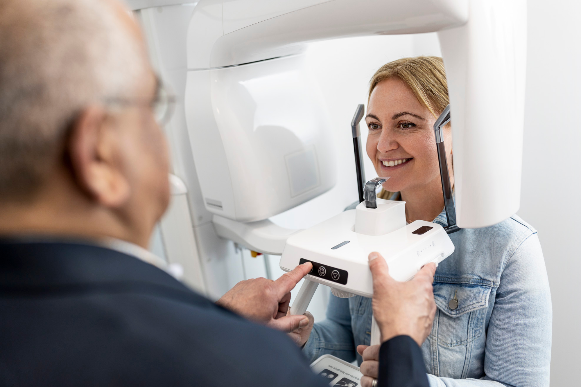
You will need to take off your glasses, hair accessories, and jewellery. You’ll be requested to stand with your chin resting on a small support for precise positioning. To stabilise your head, you will be asked to bite down on a sanitised mouthpiece.
Once your position is correct, the X-ray machine will orbit around your head, creating a panoramic image. This process is swift, and you should not experience any discomfort.
How to prepare & what to bring to your appointment?
No preparation is necessary for your dental imaging appointment. Please bring:
Your referral form - please note we accept referral forms from all providers
Medicare card
Pension or concession card
Any prior imaging
When can I expect my doctor to receive the results?
Our radiologist will analyse your scan and send the findings directly to your dentist or doctor when they are ready. Subsequently, your dentist or doctor will examine the results and provide you with an explanation.
Fees and Billing
As a radiology provider dedicated to our community, we typically bulk bill Medicare directly for most cases. However, please verify this with the clinic you are attending before your appointment.
Find a clinic
We have multiple locations across Victoria, Western Australia, and South Australia.